How Medical Imaging Helps People Beat Cancer
Cancer. The word by itself is enough to inspire fear, anger, and profound sadness. There are few Americans alive today who haven’t had their lives touched in some way by this dreaded set of diseases. That’s why it comes as such welcome news that cancer survival rates in the United States are climbing thanks to revolutionary advancements in medical imaging.
What is Medical Imaging?
Medical imaging refers to the different technologies used to view the human body in order to diagnose, monitor, or treat medical conditions. Even if you’ve never had personal experience with these medical imaging technologies, chances are you’ve heard of one or more of the following:
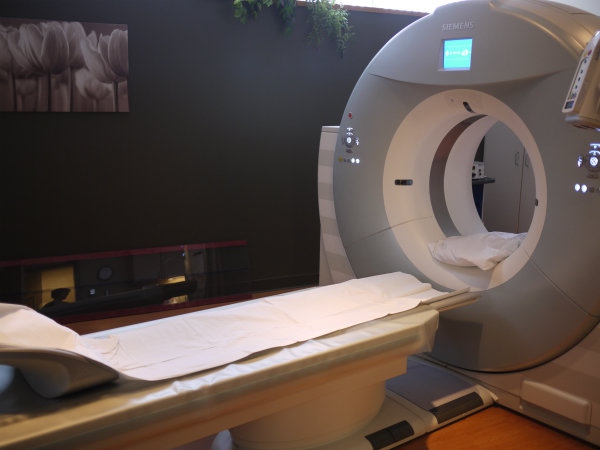
· Computed Tomography (CT)
· Magnetic Resonance Imaging (MRI)
· Positron Emission Tomography (PET)
· Ultrasound/Medical Sonography
· X-Ray
Each of these technologies provides doctors with different information about the area of the body being studied or treated, related to possible disease, injury, or the effectiveness of medical treatment.
Medical imaging has come a long way in the past 30 years. That’s why the New England Journal of Medicine ranked imaging as one of the top medical developments of the past 1,000 years. It has helped doctors develop new ways to detect, diagnose, and treat a wide variety of medical conditions.
When it comes to cancer specifically, medical imagining has helped doctors improve survival rates for their patients three key ways:
Medical Imaging Means Earlier Detection and Diagnosis
Most cancers are difficult to detect in their earliest stages, but Medical imaging helps doctors see deep within the body to find even tiny tumors. For example, Low-dose lung computed tomography (LDCT) can detect tumors as small as a grain of rice and has been shown to reduce lung cancer deaths by 20 percent compared to chest X-ray in high-risk patients.
Using medical imaging technologies to screen for colon and breast cancer has also dramatically improved rates of early detection. CT colonography, which uses a low-dose CT scan to detect polyps and tumors in the colon is faster to perform than traditional colonoscopy, and eliminates the need for sedation, making it less invasive as well.
When it comes to breast cancer detection, traditional mammography correctly identifies cancer in about 78 percent of women who have breast cancer overall, and 83 percent in women over 50. However for women whose mammogram detects an abnormality, ultrasound helps prevent unnecessary biopsies, and for women with dense breasts, who are at higher risk of developing breast cancer, magnetic resonance imaging (MRI) can detect early tumors.
Medical Imaging Helps Doctors Stage and Monitor Cancer More Effectively
Accurately staging cancer is an essential prerequisite for prescribing the most effective, individualized treatment plans. Medical imaging helps doctors because it detects disease at the cellular level, before symptoms are noticeable.
A good example is Positron emission tomography (PET), which - when combined with CT (PET/CT) - provides detailed images of internal structures, as well as signs of abnormal activity. These detailed imagines of internal structures help doctors identify and size tumors for more accurate staging.
PET/CT also help doctors plan and monitor therapy because abnormalities can develop in areas that appear normal in images taken with more traditional anatomical imagining technologies. This technology has improved survival rates in particular for patients with lymphomas, lung cancer, gynecological cancer, esophageal cancer and colorectal cancer, to name a few.
Medical Imaging Helps Doctors Treat Cancer More Precisely
One problem with treating cancer is that it can be difficult to treat with precision. The result is that healthy cells and parts of the body can suffer damage or side effects as a result of the cancer treatment itself. Medical imaging makes it easier for doctors to target tumors with greater precision, enabling them to avoid this unintended consequence of their treatment therapies.
For example, intensity-modulated radiation therapy (IMRT) uses imaging to target tumors with great precision, limiting the harm to healthy cells. Compared to standard radiotherapy, IMRT allows radiation oncologists to use stronger radiation doses since IMRT spares the surrounding healthy tissue from unnecessary exposure.
Additionally, research conducted at the Cedars-Sinai Maxine Dunitz Neurosurgical Institute’s Department of Neurosurgery is looking into ways doctors can use a new, targeted imaging agent to guide removal of malignant brain tumors and other cancers without damaging healthy tissue.
Medical imaging provides doctors with a set of powerful tools to assist with the detection, diagnostic, and treatment of cancer. Developments in these technologies have already had a dramatically positive impact on cancer survival rates, and thanks to research ongoing, we can expect that trend to continue.
Slidell Memorial Hospital is so committed to harnessing the full potential of medical imaging for our patients we offer a full range of both diagnostic and therapeutic procedures with the highest quality images.
We also recently completed a $1.5 million renovation project that enables us to provide a wide array of imaging services including Computed Tomography (CT), Interventional Mammography, Magnetic Resonance Imaging (MRI), Nuclear Medicine, Ultrasound and X-ray with Special Procedures.
Additionally, SMH's Medical Imaging department offers the only 64-slice CT scanner in Eastern St. Tammany Parish.
Call us at (985)280-8585 today to schedule an appointment with one of our Medical Imaging clinicians to learn more.
As a 229-bed acute care hospital located in the heart of Slidell, LA, Slidell Memorial Hospital provides access to cancer services, as well as the latest treatments, technology and expert physicians.